Your Back Pain is Unique, No one knows your pain better than you and understanding the mechanics will help you relieve the pain.
Your path to pain relief starts now.
You know your pain. We understand the science behind relief.
Understanding the Causes and Generators of Back Pain
Back pain is one of the most common medical complaints worldwide, affecting up to 80% of adults at some point in their lives. In clinical medicine, isolating back pain requires distinguishing between etiologies (the underlying diseases, habits, or injuries that create pathology) and pain generators (the exact anatomical structures innervated by nociceptive nerve fibers transmitting pain signals to the brain).
Tracing how these physical structures malfunction provides a clear blueprint of why the back fails and how discomfort develops.
The Primary Anatomical Pain Generators
For tissue to generate pain, it must possess sensory nerve endings (nociceptors) sensitive to mechanical deformation, inflammation, or chemical irritation. In the human spine, several distinct structures serve as primary pain generators:
1. Intervertebral Discs (Discogenic Pain)
Discs act as hydraulic shock absorbers between adjacent vertebral bodies, consisting of a gel-like center (nucleus pulposus) surrounded by a tough, fibrous ring (annulus fibrosus).
Mechanism: While the inner two-thirds of the disc are largely aneural, the outer third of the annulus fibrosus is heavily innervated by the sinuvertebral nerve.
Pathology: Micro-tears in the outer annulus allow inflammatory cytokines from the nucleus pulposus to leak out, sensitizing local nerve endings. Over time, internal disc disruption or chronic degeneration produces deep, dull axial low back pain that worsens when sitting or flexing forward.
2. Facet Joints (Zygapophysial Joint Arthropathy)
Positioned at the posterior aspect of each spinal segment, paired facet joints guide and constrain spinal motion.
Mechanism: Like the knee or hip, facet joints are synovial joints lined with cartilage and encased in a fibrous capsule innervated by the medial branch of the dorsal ramus.
Pathology: Repetitive loading, cartilage wear, or disc collapse (which shifts axial load rearward onto the facets) triggers osteoarthritis. Facet-mediated pain typically sharpens during spinal extension (arching backward), rotational twisting, or prolonged standing.
3. Nerve Roots (Radicular Pain)
Spinal nerves exit the vertebral column via lateral openings known as the neural foramina.
Mechanism: Unlike somatic back pain, which stays localized, impingement or chemical irritation of a spinal nerve root produces radiculopathy (often called sciatica).
Pathology: A herniated disc or bony spur (osteophyte) compresses the nerve root. Pain is sharp, shooting, or electric, traveling down a specific dermatome in the buttock, leg, or foot, often accompanied by numbness, tingling, or motor weakness.
4. Myofascial Structures and Ligaments
The spine is stabilized by an intricate network of deep postural muscles (multifidus, erector spinae) and robust ligaments (ligamentum flavum, interspinous ligaments).
Mechanism: Acute overload, prolonged eccentric contraction, or compensation for underlying spinal instability causes microscopic muscle fiber tearing and localized ischemia.
Pathology: Trigger points develop—hyperirritable spots within taut bands of skeletal muscle fibers—producing localized, aching pain and protective muscle spasms that restrict movement.
5. Sacroiliac (SI) Joint
Located at the base of the spine where the sacrum connects to the iliac bones of the pelvis, the SI joint transfers load between the upper body and legs.
Mechanism: Innervated by sacral nerve branches, this joint is susceptible to mechanical shear, ligamentous laxity (common during pregnancy), and inflammatory sacroiliitis.
Pathology: Pain typically localizes to the posterior superior iliac spine (the “dimple” of the lower back) and can refer pain into the groin, buttock, and posterior thigh.
The Nervous System: Acute vs. Nociplastic Pain
Not all back pain correlates neatly with structural damage on an MRI. Many individuals with prominent disc bulges report no pain, while others with “clean” imaging experience severe disability.
Acute Nociceptive Pain: Operates as a direct alarm system in response to tissue threat or inflammation. Healing generally follows a predictable biological timeline over 6 to 12 weeks.
Central Sensitization (Nociplastic Pain): If pain persists, the central nervous system can undergo neuroplastic remodeling. The dorsal horn of the spinal cord and cortical pain-processing networks become hyper-responsive. The volume knob of the nervous system is turned up, meaning normal mechanical pressure, minor stress, sleep deprivation, or emotional distress can trigger severe spinal pain without ongoing tissue damage.
Resolving back pain effectively relies on moving beyond vague diagnoses of “a bad back” to identify the precise tissue generators involved, while accounting for the mechanical loads, systemic health, and neural sensitivity that sustain them.
Am I a candidate for artificial disc replacement?
Lumbar Total Disc Replacement (lTDR)
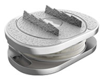
Lumbar total disc replacement replaces a degenerated lumbar disc with an articulating prosthesis to treat discogenic lower back pain. Ideal candidates have single- or two-level degenerative disc disease; severe facet arthrosis, spondylolisthesis, and osteoporosis are contraindicated. Mechanically, lTDR preserves segmental motion and lordosis, preventing fusion-induced stress transfer across adjacent kinetic chain levels.
Key Clinical Breakdown:
- Procedure Definition: Anterior-approach total reconstruction of the intervertebral disc utilizing a mobile- or fixed-bearing prosthetic implant to restore disc height and neural foramen patency.
- Qualifying Indications: Severe, disabling single-level or contiguous two-level discogenic low back pain confirmed by diagnostic imaging and conservative care failure for at least six months.
- Contraindications: High-grade spondylolisthesis, severe facet joint osteoarthritis, central canal spinal stenosis requiring extensive posterior decompression, severe osteopenia/osteoporosis, or major vascular anatomic anomalies.
- Biomechanical Advantage: Re-establishes normal disc height and segmental lordosis while retaining mobility, avoiding the rigid fusion biomechanics that accelerate adjacent segment degeneration in the lower lumbar spine.
Dr. Karsten Ritter-Lang a top orthopedic spine surgeon, explains how this occurs. Fuse one level, and when the level next to it wears down, that needs surgery as well, and the cycle continues. This is how you end up in a spinal fusion cascade. This is one reason motion-preserving options matter so much. The goal isn’t exclusively to fix the level causing pain today. It’s to hopefully avoid turning one treated level into several surgeries.
If you’re dealing with chronic leg and/or back pain and considering spine surgery, it’s worth asking whether a motion-preserving option is available to you from the start, and if fusion is the recommendation, why that’s the case. If you’ve already had a spine procedure and you’re facing a second surgery, the question becomes whether that next level could be treated in a way that preserves mobility and function.
Patients Seek Artificial Disc Replacement in Germany
International patients frequently travel to specialized spine centers in Germany to access advanced motion-preserving spine surgery alternatives to conventional spinal fusion.
Key clinical differentiators include:
- Multi-Level Approaches: Routine performance of multi-level lumbar (L4-L5, L5-S1) and cervical disc arthroplasty.
- Prosthetic Technology: Access to next-generation viscoelastic and unconstrained disc prosthetics (e.g., M6-L, M6-C, ProDisc-L, LP-ESP, activL).
- Anterior Retroperitoneal Exposure: Muscle-sparing anterior lumbar approaches designed to preserve posterior musculature and reduce adjacent segment disease (ASD).
About Dr. Karsten Ritter-Lang: Motion Preservation & Spine Arthroplasty
Dr. Karsten Ritter-Lang is recognized internationally for his clinical focus on intervertebral disc prosthetics and complex spinal reconstruction.
- Surgical Experience: Over 30 years dedicated to motion-preserving spinal procedures, having performed thousands of cervical and lumbar artificial disc replacements.
- Academic Lineage: Trained directly under Prof. Dr. Kurt Schellnack and Dr. Karin Büttner-Janz at the Charité University Hospital in Berlin—the pioneering research team that developed the original Charité artificial disc prosthesis.
- Clinical Focus: Multi-level lumbar disc replacement, motion-preserving reconstruction for degenerative disc disease, and alternatives to spinal fusion.
Remote Consultations & International MRI Reviews
For patients seeking preliminary assessments or second opinions regarding total disc replacement and spinal stenosis treatments:
- Better Disc Replacement: Detailed comparative guides on motion preservation vs. spinal fusion, clinical indications for lumbar total disc replacement (TDR), and multi-level case analyses.
BetterDiscReplacement
- Stenum Hospital International Patient Portal: Preliminary remote MRI evaluations, international intake protocols, and surgical technology overviews for European and overseas patients
Stenum Hospital