A surgical consultation for spinal fusion should never be a one-way lecture. When a surgeon recommends permanently locking vertebrae together with titanium screws, rods, and bone graft, the biomechanics of your entire spine are fundamentally altered.
Generalized statistics in medical brochures mean very little compared to what happens in your specific surgeon’s operating room. To protect your long-term mobility and avoid becoming trapped in a cycle of repeat operations, three critical areas require direct interrogation: Adjacent Segment Disease (ASD), true fusion rate benchmarks, and their exact clinical protocol if hardware nonunion (pseudarthrosis) occurs.
Here is the exact framework of questions to bring to your surgical consultation.
Category 1: Adjacent Segment Disease (ASD) Rates and Biomechanical Protection
When a spinal segment is fused, it turns two or more flexible vertebrae into a single rigid lever arm. The mobile discs directly above and below that fusion now absorb abnormal torsional stress and load, frequently accelerating disc herniation, facet arthropathy, and stenosis.
“What is your personal rate of symptomatic Adjacent Segment Disease requiring reoperation at 2, 5, and 10 years?”
Why ask: Published literature documents that 2% to 4% of fusion patients develop symptomatic ASD per year, with roughly 20% to 30% showing radiographic or symptomatic breakdown within a decade. You need to know if your surgeon tracks their own reoperation rate or merely quotes national averages.
“How does the health of my adjacent discs look right now on MRI?”
Why ask: If an adjacent disc already shows mild dehydration (black disc), modic changes, or bulging, fusing the neighboring segment acts as an accelerant. If an adjacent level is already compromised, you must ask how they plan to avoid triggering rapid collapse.
“How do you plan to preserve sagittal balance and lordosis during this fusion?”
Why ask: Flatback deformity or failing to restore natural lumbar or cervical lordosis drastically increases mechanical sheer stresses at neighboring levels. Your surgeon should be able to explain how cage angle and contouring protect adjacent levels.
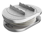
“Am I a candidate for motion-preserving technology—such as artificial disc replacement (ADR) or a hybrid construct—instead of a pure fusion?”
Why ask: If the problem is discogenic pain or radiculopathy without severe instability, motion preservation avoids creating the rigid lever arm that causes ASD in the first place.
Category 2: Fusion Rate Benchmarks and Biological Reality
A spinal fusion is essentially an attempt to trick the body into healing an intentional fracture. Hardware does not heal you; bone bridging across the disc space or transverse processes does.
“What is your verified radiographic fusion rate at 12 months for this specific approach?”
Why ask: Fusion benchmarks vary significantly depending on technique (e.g., ACDF vs. ALIF vs. TLIF vs. posterolateral fusion without interbody support) and the number of levels. Single-level procedures often achieve 90% to 95% fusion rates, but multi-level procedures can drop significantly.
“How do you define a ‘successful fusion’ in your practice?”
Why ask: There is a major clinical distinction between radiographic fusion (solid bone bridging visible on dynamic imaging) and clinical success (meaningful pain relief). Hardware can stay in place while bone fails to grow, or bone can fuse solidly while nerve pain persists. Demand clarity on both.
“What exact biologic or graft material do you use to achieve fusion?”
Why ask: Autograft (your own bone), allograft (cadaver bone), synthetic ceramics, and bone morphogenetic proteins (rhBMP-2) carry vastly different fusion success rates and risk profiles. Ask why they prefer their specific biologic choice for your bone quality.
“How do you objectively verify solid fusion post-operatively?”
Why ask: Plain flexion-extension X-rays often miss micro-motion or pseudarthrosis around pedicle screws. Find out if they routinely order fine-cut CT scans at 6 or 12 months to confirm continuous trabecular bone bridging before clearing you for unrestricted physical activities.
Category 3: Protocol for Hardware Nonunion (Pseudarthrosis) and Failure
If bone fails to knit within 6 to 12 months, the condition is classified as pseudarthrosis (nonunion). Because metal cannot withstand repetitive cycling indefinitely without biological bone support, the hardware eventually loosens, halos, or snaps under metal fatigue. You need to know their contingency plan before the first incision is made.
“What is your precise protocol if I have persistent or worsening pain at the 6-to-12-month mark?”
Why ask: A surgeon should never dismiss ongoing pain as “normal scar tissue.” You need an established escalation pathway: dynamic X-rays, thin-slice CT reconstructions, diagnostic facet/hardware blocks, or nuclear medicine SPECT-CT scans to isolate motion.
“What conservative steps do you exhaust before considering revision surgery for nonunion?”
Why ask: In asymptomatic or minimally symptomatic delayed unions, surgeons often employ pulsed electromagnetic field (PEMF) bone growth stimulators, metabolic bone workups, or activity restrictions before jumping back into the OR.
“If revision surgery is required for nonunion, what is your approach?”
Why ask: A successful revision requires changing the biomechanical environment. If a posterior approach (like a TLIF) failed to fuse, a skilled surgeon will often propose revising from the anterior or lateral column (ALIF/LLIF) to achieve a larger footprint and better compressive loading, rather than simply putting larger screws down the same compromised bone channels.
“What are my individual biological risk factors for pseudarthrosis, and how are we mitigating them prior to surgery?”
Why ask: Bone density (osteopenia/osteoporosis), Vitamin D levels, diabetes, systemic inflammation, and NSAID use directly suppress osteogenesis. If your surgeon does not order a pre-op DEXA scan or Vitamin D panel for major reconstructive fusion, they are leaving biological variables to chance.
Consultation Action Checklist
Request a written breakdown of the planned surgical approach (anterior, posterior, transforaminal, or lateral) and why it yields the highest fusion rate for your anatomy.
Clarify all implant materials: titanium, PEEK, or porous 3D-printed titanium designed for cellular bone in-growth.
Require explicit clearance protocols detailing when lifting restrictions are removed, verified by imaging rather than an arbitrary calendar date.
Ensure a second opinion is obtained, preferably from a motion-preservation specialist, to confirm fusion is an absolute clinical necessity rather than an automatic institutional habit.
Medically Reviewed By
Dr. med. Karsten Ritter-Lang M.D.
Diploma of medicine (Institut of Immunology Charité)
1988 – 1991
Doctorade of medicine (Institut of Immunology Charité)
Senior consultant of spinal surgery in two hospitals (DRK Berlin Westend and Stenum Hospital) and private practice in Potsdam,ENDOCERT Certified awarded the title and certification of Specialist for Trauma Surgery (Facharzt für Unfallchirurgie) in 2009
Schedule Surgery Now
Go to
Investigated multi-axis kinematics and coupled physiological motion of viscoelastic artificial cervical discs (M6-C) compared to intact segments.
Citation: Patwardhan AG, Tzermiadianos MN, Tsitsopoulos PP, Voronov LI, Renner SM, Reo ML, Carandang G, Ritter-Lang K, Havey RM. Primary and coupled motions after cervical total disc replacement using a compressible six-degree-of-freedom prosthesis. European Spine Journal, 2012; 21(4): 618–629.
Cervical and Lumbar Disc Arthroplasty: A Review of Current Implant Design and Outcomes – PMC
Compared safety profiles, range of motion (ROM), pain reduction (VAS), and disability scores (ODI) between single-level versus multi-level lumbar artificial disc replacements.
Citation: Schätz C, Ritter-Lang K, Gössel L, et al. Comparison of Single-Level and Multiple-Level Outcomes of Total Disc Arthroplasty: 24-Month Results. International Journal of Spine Surgery (IJSS), 2015; 9: 14
Long-term outcomes following lumbar total disc replacement with M6-L – PMC
Evaluated 24-month clinical outcomes, restoration of intervertebral disc height, and segment lordosis in lumbar degenerative disc disease treated with the M6-L prosthesis.