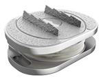
Intervertebral discs act as hydraulic shock absorbers between the vertebrae, composed of a tough, fibrous outer ring (the annulus fibrosus) and a soft, gel-like core (the nucleus pulposus). Over time, mechanical stress, aging, dehydration, or sudden trauma can alter their structure.
The 7 TYPES OF DISC PROBLEMS
From Normal Disc to Advanced Disc Degeneration
1. Disc Degeneration (Internal Desiccation & Wear)
The earliest phase of breakdown occurs at the cellular and biochemical level as discs lose hydration and proteoglycan content.
Key Characteristics:
Reduced disc height: Loss of fluid volume reduces vertical clearance between vertebrae.
Desiccation on imaging: Appears noticeably darker (“black disc”) on T2-weighted MRI due to water loss.
Surface irregularities: Micro-tears begin developing in the inner layers of the annulus.
Mild biomechanical changes: Shock absorption declines, shifting uneven loads onto the facet joints.
2. Disc Bulge (Generalized Circumferential Extension)
A bulge occurs when the outer fibrous ring weakens uniformly, causing the disc margin to extend past its anatomical perimeter across more than 25% (often 50% or more) of the disc’s circumference.
Key Characteristics:
Broad-based extension: The disc protrudes uniformly beyond the edges of the adjacent vertebral endplates.
Intact annulus: The outer fibers remain structurally continuous; no focal rupture has occurred.
Mild degenerative wear: Typically an age-related adaptation to axial loading, often asymptomatic unless it narrows the spinal canal or neuroforamen.
3. Disc Protrusion (Focal Herniation – Contained)
A protrusion represents the earliest subtype of true disc herniation. Instead of a broad, symmetrical bulge, the nucleus pushes focally into a localized defect in the annulus (covering less than 25% of the disc circumference).
Key Characteristics:
Focal outward push: The base of the displaced disc material against the parent disc remains wider than the distance it extends outward.
Contained displacement: The outer lamellae of the annulus fibrosus or the posterior longitudinal ligament (PLL) still enclose the displaced gel.
Asymmetrical contours: Can produce unilateral nerve irritation depending on whether the protrusion is central, paracentral, or foraminal.
4. Disc Extrusion (Uncontained Rupture)
An extrusion occurs when the nucleus pulposus completely tears through all layers of the annulus fibrosus, breaking beyond the posterior longitudinal ligament.
Key Characteristics:
Narrow neck, wider head: The dome of displaced disc tissue is wider in at least one dimension than the narrow “stalk” or aperture connecting it back to the central disc space.
Uncontained material: Nuclear gel spills into the epidural space.
Acute inflammatory response: Exposure of the vascularized epidural space to foreign nuclear proteins triggers a strong chemical and immune-mediated inflammatory reaction.
5. Sequestered Disc (Free Fragment)
Also known as a “free fragment,” sequestration is an advanced form of extrusion where a segment of the extruded nuclear material breaks completely free from the parent disc.
Key Characteristics:
Loss of continuity: The extruded piece has no remaining tissue bridge connecting it to the original intervertebral disc space.
Migration potential: The loose fragment can migrate cranially (upward) or caudally (downward) along the anterior or posterior epidural space.
Severe mechanical blockage: Frequently causes acute, sharp radicular pain or local canal compromise, though free fragments often trigger an enzymatic reaction that may resorb over time.
6. Disc Herniation with Nerve Compression (Radiculopathy)
While “herniation” encompasses protrusions, extrusions, and sequestrations, clinically significant herniation refers to displaced disc material directly compressing, displacing, or chemically irritating adjacent neural structures.
Key Characteristics:
Direct neural impingement: Displaced material contacts or flattens the traversing nerve root, exiting nerve root, or thecal sac.
Classic radicular symptoms: Causes dermatomal pain (e.g., sciatica), numbness, tingling, diminished reflexes, or motor weakness along the path of the compressed nerve.
Dual insult mechanism: Symptoms stem from both direct physical mass-effect (ischemia from pressure) and neurochemical inflammation caused by cytokines leaked from the nucleus pulposus.
7. Disc Collapse (Advanced Degenerative Disc Disease)
The end stage of chronic disc degeneration, where the intervertebral cushion is almost entirely lost.
Key Characteristics:
- Severely obliterated disc space: The disc is flattened to a thin, fibrotic scar tissue strip.
- Close approximation of vertebral bodies: Opposing vertebral endplates sit almost in direct contact.
- Secondary osseous remodeling: Significant subchondral sclerosis (bone hardening) and large peripheral bone spurs (osteophytes) form to stabilize the unstable segment.
- Foraminal stenosis: Severe height loss narrows the neural exit canals, often causing chronic structural nerve entrapment even in the absence of a soft herniation.
Am I a candidate for Disc Replacement Surgery?
Disc replacement surgery provides a less invasive alternative to spinal fusion surgery and protects against adjacent level degeneration.
“Advanced disc replacement solutions have allowed us to achieve a success rate of 99%”
Dr. Karsten-Ritter-Lang Disc Replacement Surgeon
“After years of suffering, I avoided spinal fusion surgery, had Disc Replacement Surgery, went to Dr. Ritter-Lang and had a wonderful result.”
“Dr. Ritter-Lang gave me a solution that I was not being offered by my doctors in the US”
Medically Reviewed By
Dr. med. Karsten Ritter-Lang M.D.
Diploma of medicine (Institut of Immunology Charité)
1988 – 1991
Doctorade of medicine (Institut of Immunology Charité)
Senior consultant of spinal surgery in two hospitals (DRK Berlin Westend and Stenum Hospital) and private practice in Potsdam,ENDOCERT Certified awarded the title and certification of Specialist for Trauma Surgery (Facharzt für Unfallchirurgie) in 2009