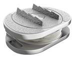
The recovery timeline and restriction differences between Lumbar Spinal Fusion vs. Lumbar Artificial Disc Replacement
Recovery, Timelines, and Restrictions
Lumbar spinal fusion (arthrodesis) and lumbar artificial disc replacement (ADR / arthroplasty) represent two distinct surgical paradigms for treating severe degenerative disc disease and discogenic low back pain. Because their primary mechanical goals are opposites—fusion aims to eliminate motion entirely to achieve solid osseous union, while ADR aims to preserve physiologic kinematics and shock absorption—their postoperative protocols, healing dynamics, recovery milestones, and physical restrictions diverge significantly.
Comparative Overview of Recovery Milestones
The standard clinical milestones and phases of rehabilitation for both procedures under uncomplicated circumstances compare as follows:
Hospital Stay
- Lumbar Spinal Fusion: Typically 2 to 4 days (longer for open posterior approaches requiring extensive paraspinal muscle dissection).
- Lumbar Disc Replacement: Typically 1 to 2 days (the anterior retroperitoneal approach spares posterior musculature and allows rapid inpatient recovery).
Initial Mobilization
- Lumbar Spinal Fusion: Walking within 24 hours under strict log-rolling and spinal precautions.
- Lumbar Disc Replacement: Walking the same day or within 12 to 24 hours; early controlled movement is actively encouraged to restore joint kinematics.
Sedentary / Desk Work
- Lumbar Spinal Fusion: Return after 4 to 6 weeks, requiring frequent posture changes and ergonomic workstation modifications.
- Lumbar Disc Replacement: Return after 2 to 3 weeks, as sitting tolerance improves rapidly without rigid fixation constraints.
Physical Therapy Initiation
- Lumbar Spinal Fusion: Delayed until 6 to 12 weeks post-operatively to await initial radiographic evidence of bridging bone callus.
- Lumbar Disc Replacement: Early initiation at 2 to 4 weeks, focusing on active core engagement, pelvic stabilization, and range of motion restoration.
Biological Integration / Fusion
- Lumbar Spinal Fusion: Requires 6 to 12 months for solid trabecular bone bridging across the interbody space and posterior elements.
- Lumbar Disc Replacement: Requires 6 to 12 weeks for osteointegration and biological bone ingrowth into porous titanium or hydroxyapatite-coated endplates.
Unrestricted / Impact Activity
- Lumbar Spinal Fusion: Cleared at 6 to 12+ months, strictly contingent upon radiographic confirmation of solid bony union.
- Lumbar Disc Replacement: Cleared at 3 to 6 months once dynamic muscular control, core strength, and surrounding soft tissues adapt.
Post-Operative Restrictions and Protocol Comparison
Lumbar Spinal Fusion Protocols
- The “BLT” Ban (No Bending, Lifting, Twisting): Patients must adhere to strict activity restrictions for the first 3 months. Bending the lumbar spine and rotational torso twisting are prohibited to protect graft containment and pedicle screw stability. Lifting is strictly capped at 5 to 10 lbs initially.
- External Orthosis / Bracing: Rigid or semi-rigid lumbar braces (such as a TLSO or LSO) are frequently prescribed for 6 to 12 weeks to minimize micro-motion across the fusion bed and avoid mechanical shear on stabilizing hardware.
- NSAID Contraindication: Non-steroidal anti-inflammatory drugs (NSAIDs such as ibuprofen, naproxen, and meloxicam) are strictly contraindicated for 3 to 6 months post-operatively. NSAIDs inhibit COX enzymes and suppress osteoblast activity, significantly increasing the risk of non-union (pseudarthrosis).
- Postural Restrictions: Prolonged sitting (beyond 20–30 consecutive minutes) is discouraged in the early weeks to avoid excessive axial compressive loading on the healing graft.
- Athletic Restrictions: Rotational sports (golf, tennis), high-impact conditioning (running), and heavy resistance training are prohibited until radiographic confirmation demonstrates definitive bridging bone (typically 6–12 months).
Lumbar Artificial Disc Replacement Protocols
- Encouraged Early Mobility: Because primary mechanical stability is established immediately upon insertion via endplate keels, spikes, or teeth, natural walking and gentle, controlled extension/flexion are encouraged early to maintain muscular tone and joint flexibility.
- Progressive Weight Lifting Limits: Lifting limits begin at 10 to 15 lbs during weeks 1 to 4, advancing steadily to 30+ lbs by weeks 6 to 8 as abdominal and lumbar stabilizers recover.
- Minimal External Immobilization: Rigid braces are generally avoided because immobilization can lead to facet joint stiffness and muscular atrophy. Soft elastic corsets are only used optionally for comfort during prolonged upright activities in the first 2 to 3 weeks.
- Pharmacological Flexibility: Short courses of NSAIDs may be utilized if necessary for pain management or periprocedural inflammation, as implant integration relies on localized bone ongrowth/ingrowth rather than large-scale de novo bone mass synthesis across the disc space.
- Accelerated Return to Sport: Low-impact cardio (recumbent bike, elliptical, swimming) begins at 4 to 6 weeks. Rotational and athletic activities (such as golf, skiing, and tennis) typically resume by 3 to 4 months once dynamic core stability is restored.
Underlying Biomechanical and Healing Differences
Healing Biology: Bone Bridge vs. Osseointegration
In spinal fusion, surgery initiates a race between biological bone healing and mechanical hardware fatigue. The internal fixation (screws, rods, cages) provides only temporary stabilization; if solid bridging bone does not form across the vertebral endplates within 6 to 12 months, the cyclic loading will eventually lead to screw loosening, rod fracture, or pseudoarthrosis. In contrast, artificial disc replacement relies on immediate press-fit mechanical fixation followed by microscopic osteointegration, where host bone cells anchor directly into porous titanium plasma spray or hydroxyapatite coatings over 6 to 12 weeks.
Kinematics and Adjacent Segment Mechanics
A successful lumbar fusion eliminates the 4 to 10 degrees of sagittal and coronal motion at the index level. This rigid lever arm forces the adjacent mobile segments (most commonly L4-L5 above an L5-S1 fusion) to absorb disproportionate hypermobility, intradiscal pressure, and shear stresses. Over time, this acceleration of wear manifests as Adjacent Segment Disease (ASD). Conversely, lumbar disc arthroplasty aims to maintain physiologic range of motion, center of rotation, and lordotic alignment, thereby dampening mechanical stress transfer to adjacent functional spinal units.
Surgical Approach and Soft Tissue Preservation
While modern minimally invasive fusion techniques exist, many posterior or transforaminal fusions require extensive retraction and electrocautery of the multifidus and erector spinae musculature, which can lead to localized denervation and persistent muscular stiffness. Total disc replacement is predominantly performed via an anterior retroperitoneal approach, traversing natural anatomical tissue planes without cutting or stripping the posterior paraspinal musculature.
Remote Consultations & International MRI Reviews
For patients seeking preliminary assessments or second opinions regarding total disc replacement and spinal stenosis treatments:
- Better Disc Replacement: Detailed comparative guides on motion preservation vs. spinal fusion, clinical indications for lumbar total disc replacement (TDR), and multi-level case analyses.
BetterDiscReplacement